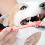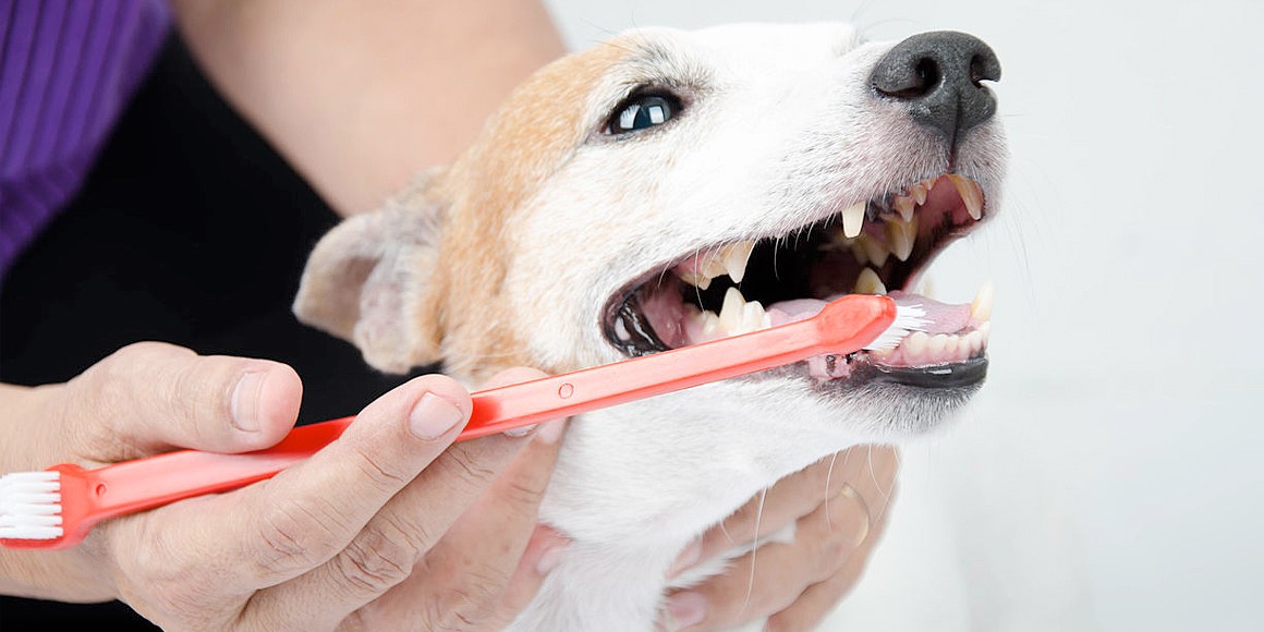
Veterinary dental radiographs (X-rays) are a vital diagnostic tool that allow us to see below the gum line—where over 60% of dental disease occurs. Without them, many painful conditions in your pet’s mouth may go unnoticed and untreated.
What Are Dental Radiographs?
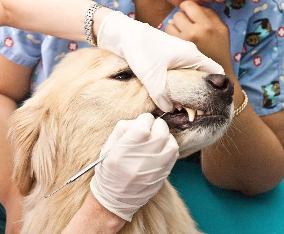
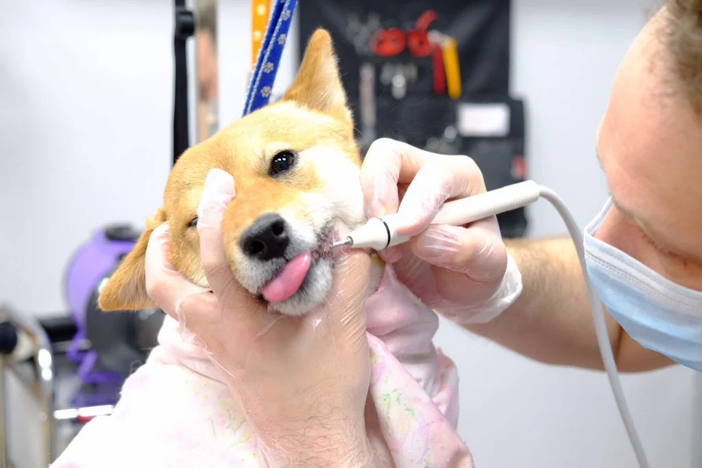
Dental radiographs are high-resolution X-rays of the teeth and jaw structures. They’re taken while your pet is under general anaesthesia during a dental procedure. These images allow us to assess:
- Tooth roots
- Bone levels
- Periodontal pockets
- Abscesses or cysts
- Resorptive lesions
- Unerupted or missing teeth
- Jaw fractures
Why They’re Essential in Pet Dentistry
- Detect Hidden Disease
Problems like root infections, fractured tooth roots, or bone loss can be missed during visual exams. Radiographs provide a complete view. - Guide Treatment Decisions
Dental X-rays help determine whether a tooth can be saved, restored, or needs extraction. This ensures more targeted, effective care. - Prevent Future Complications
Early detection of resorptive lesions or impacted teeth helps prevent pain and future dental surgery. - Support Accurate Diagnosis of Masses or Cysts
When oral masses are present, radiographs can reveal bone involvement or identify associated dentigerous cysts. - Assess Treatment Success
Post-op radiographs confirm successful extractions and root canal procedures, and identify any remaining root fragments or complications.
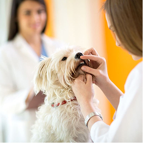
Standard of Care
At Melbourne Animal Dentistry, full-mouth dental radiographs are standard in all COHATs and dental procedures. This commitment to diagnostic excellence ensures we never miss a hidden issue, and your pet receives the most appropriate and effective care.
Book a Consultation
It’s easy and free!